THE HUMAN EYE
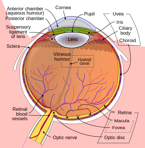
Our human eye is a complex organ with a variety of layers and muscles. Obviously, it is used to help humans use thier sense of sight.The human eye is unique in that it accommodates for different light intensities and also focuses light rays that come from different distances from the eye. Light is then converted into impulses and sent to the brain where this image is perceived as what it looks like. Vision is very dependent on light. The light rays are reflected off object and then make their way into the eye. The light rays enter the eye through the cornea and the cornea refracts the light as it enters the eye. After passing through the cornea the light rays pass through the opening called the pupil. The iris is the part of the eye that controls the dilation of the pupil. This allows a certain amount of light into the eye. However, many humans experience vision problems that either impair their vision or prevent them from seeing altogether. At 1 inch (2.54 cm) wide, 1 inch deep and 0.9 inches (2.3 cm) tall, the human eye is composed of fifteen main parts. Maintaining the shape of the eye is the tough, outermost layer the sclera. A clear layer, the cornea, covers the front of the sclera. Light first passes through the cornea when it enters the eye. Attached to the sclera are the extraocular muscles that move the eye. The choroid is the second layer of the eye. It contains the blood vessels that supply blood to all structures of the eye. The choroid is composed of the ciliary body and the iris. A muscular area that is attached to the lens, the ciliary body contracts and relaxes to control the size of the lens for focusing. Functioning to color the eye, the iris is an adjustable diaphragm surrounding the pupil. It has two muscles: the dilator and the sphincter. Both control the amount of light let into the eye by adjusting the pupil size. The color of the iris is determined by the color of the connective tissue and pigment cells. The innermost layer is the retina -- the light-sensing portion of the eye. It contains rod cells, which are responsible for vision in low light, and cone cells, which are responsible for color vision and detail. In the back of the eye, in the center of the retina, is the macula. In the center of the macula is an area called the fovea centralis. This area contains only cones and is responsible for seeing fine detail clearly. Inside the eyeball there are two fluid-filled sections separated by the lens, a clear structure used to fine-tune vision. The larger, back section contains a clear, gel-like material called vitreous humor. The smaller, front section contains a clear, watery material called aqueous humor. When drainage of the aqueous humor is blocked, a disease called glaucoma can result. The eye is also unique in that it is able to move in many directions to maximize the field of vision, yet is protected from injury by a bony cavity called the orbital cavity. The eye is embedded in fat, which provides some cushioning. The eyelids protect the eye by blinking. Eyelashes and eyebrows protect the eye from particles that may injure it.There are six muscles attached to the sclera that control the movements of the eye. They are shown here with their descriptions:MusclePrimary FunctionMedial rectusmoves eye towards noseLateral rectusmoves eye away from noseSuperior rectusraises eyeInferior rectuslowers eyeSuperior obliquerotates eyeInferior obliquerotates eyeAfter passing through the cornea, light passes through the aqueous humor, lens and vitreous humor. Ultimately it reaches the retina, which is the light-sensing structure of the eye. The retina is made up of cones and rods and is lined with black pigment called melanin to lessen the amount of reflection. The retina has a central area, called the macula that is responsible for sharp, detailed vision. The color-responsive chemicals in the cones are called cone pigments and are very similar to the chemicals in the rods. Each cone cell has a red-sensitive pigment, a green-sensitive pigment, and a blue-sensitive pigment. The presence of these pigments allow the eye to be sensitive to that color. The human eye can sense almost any gradation of color when red, green and blue are mixed.In the diagram above, the wavelengths of the three types of cones (red, green and blue) are shown. The peak absorbency of blue-sensitive pigment is 445 nanometers, for green-sensitive pigment it is 535 nanometers, and for red-sensitive pigment it is 570 nanometers. Color blindness is the inability to differentiate between different colors. The most common type is red-green color blindness. This occurs in 8 percent of males and 0.4 percent of females. It occurs when either the red or green cones are not present or not functioning properly. People with this problem are not completely unable to see red or green, but often confuse the two colors. Another vision problem is vitamin A deficiency. When severe vitamin A deficiency is present, then night blindness occurs. This is when the levels of light-sensitive molecules are low due to vitamin A deficiency, there may not be enough light at night to permit vision. During daylight, there is enough light stimulation to produce vision despite low levels of retinal. Refraction is when light rays reach an angulated surface of a different material and the light rays bend. When light reaches a convex lens, the light rays bend toward the center:When light rays reach a concave lens, the light rays bend away from the center:Vision or visual acuity is tested by reading a Snellen eye chart at a distance of 20 feet. By looking at lots of people, eye doctors have decided what a "normal" human being should be able to see when standing 20 feet away from an eye chart. 20/20 vision means when standing 20 feet away from the chart you can see what a "normal" human being can see and have normal vision. If you have 20/40 vision, it means that when you stand 20 feet away from the chart you can only see what a normal human can see when standing 40 feet from the chart. 20/200 is the cutoff for legal blindness in the United States. It is also possible to have vision better than normal. A person with 20/10 vision can see at 20 feet what a normal person can see when standing 10 feet away from the chart. Hawks, owls and other birds of prey have much more acute vision than humans. A hawk has a much smaller eye than a human being but has lots of sensors (cones) packed into that space. This gives a hawk vision that is eight times more acute than a human's. A hawk might have 20/2 vision!Normally, your eye can focus an image exactly on the retina:Nearsightedness and farsightedness occur when the focusing is not perfect. When nearsightedness is present, a person can clearly see near objects, while experiencing difficulty seeing far objects. Light rays become focused in front of the retina. This is caused by an eyeball that is too long, or a lens system that has too much power to focus. Nearsightedness is corrected with a concave lens. This lens causes the light to diverge slightly before it reaches the eye, as seen here:When farsightedness is present, a person can clearly see far objects, but has trouble seeing seeing near objects. Light rays become focused behind the retina. This is caused by an eyeball that is too short, or by a lens system that has too little focusing power. This is corrected with a convex lens, as seen here:As stated earlier, to be legally blind visual acuity must be less than 20/200 with corrective lenses. Some causes of blindness include cataracts, glaucoma, macular degeneration, trauma, vitamin A deficiency, tumors, strokes, neurological diseases, hereditary diseases and toxins.The human eye is an interesting organ full of potential complications and great physics lessons.
1 comment:
does anyone else reckon that eye diagram looks like a table-tennis bat??
Post a Comment